Lymph node metastasis in patients with oral squamous cell carcinoma
OVERVIEW
This study aimed to investigate the factors related to lymph node metastasis (LNM) in patients with oral squamous cell carcinoma (OSCC). Here we share the analysis methods of bulk, single-cell, and spatial analyses to understand the landscape of the tumor microenvironment (TME) of OSCC.
We are going to update this website whenever we add data analysis continually. Please let us know if there is anything you are interested in that is not covered in our paper.
UPDATES
- 09-04-2025
- Our paper has been officially published in PLOS Genetics! 🎉
- The “How to Cite” and “Data Availability” sections have been updated with the final publication details.
- 11-01-2023
- 10-10-2022
- Updated the integration cluster analysis and added the bootstrap method.
- 08-07-2022
- Added the interactive table of cell-type-specific marker genes from TME of OSCC.
- 07-29-2022
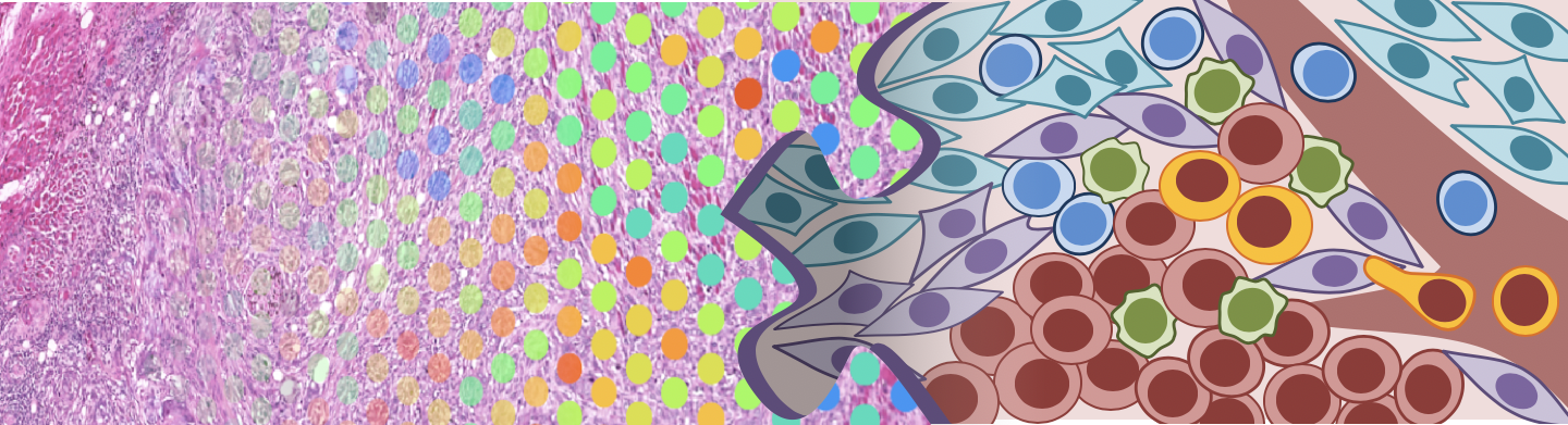
- Updated the website header image (created by co-author Shuya Kasai).
- This website is as now available to the public.
- 07-10-2022
- Edited the Reference.
- 06-20-2022
- Added spatial annotation and spatial gene expression.
- 05-08-2022
- Added the metascape results of Bulk RNA-seq and scRNA-seq.
- 05-01-2022
- 12-29-2021
- Started setting this website up.
- Started setting this website up.
Media Coverage
We are honored that our work has been featured in an official press release from The University of Texas MD Anderson Cancer Center.
- MD Anderson Newsroom: Researchers identify predictive biomarkers for oral cancer metastasis
Data Analysis Software
R (v3.6.29)
ggplot2 (v3.3.5)
tidyverse (v1.3.1)
tidyr (v1.1.3)
tibble (v3.1.4)
dplyrr (v1.0.7)
purrr (v0.3.4)
stringr (v1.4.0)

Python (v3.7.12)
Scanpy (v1.8.2)
CellChat (v1.4.0)
leidenalg (v0.8.8)
lifelines (v0.27.8)
Squidpy (v1.1.2)
matplotlib (v3.2.2)
seaborn (v0.11.2)
numpy (v1.21.5)
pandas (v1.3.5)
anndata (v0.7.8)
stlearn (v0.3.1)
scvi-tools (v0.14.5)

Additional software included
gdc-client (v1.6.0).
Metascape (v3.5),
ESTIMATE (v1.0.13),
CIBERSORTx (2022),
inferCNV (v1.11.1),
G*Power (v3.1.9.7),
Space Ranger (v1.3.0, 10x Genomics),
and Loupe Browser (v6.0.0, 10x Genomics),
NAVIGATION
We used no unpublished codes in this study. Please take a look at the individual software guides for more information. Our paper provides analysis methods. All other data supporting the results of this study are available from the corresponding author upon reasonable request.
- Metascape was used to perform DEGs enrichment analysis and protein-protein interaction analysis (https://metascape.org/).
- ESTIMATE was used to calculate the score of the purity of tumor tissue and stromal cell presence and the level of immune cell infiltration (https://bioinformatics.mdanderson.org/estimate).
- CIBERSORTx was used to estimate the abundance of cell types in the oral cancer microenvironment (https://cibersortx.stanford.edu).
- Scanpy was used for quality control and visualization of single-cell data (https://github.com/scverse/scanpy).
- CellChat was implemented to infer intercellular communications (https://github.com/sqjin/CellChat).
- The spatial transcriptome data were processed using Space ranger v1.3.0 with reference genome GRCh38 (https://www.10xgenomics.com/) .
- Spatial annotation and spatial gene expression.
- Tangram was used to map the cells within each spot (https://github.com/broadinstitute/Tangram).
- FFPE spatial deconvolution analysis
- Cell-type-specific marker genes table
- IHC staining can be found on the figshare (DOI: https://doi.org/10.6084/m9.figshare.20407344.v1)
- Statistical test of IHC analysis
- Squidpy was estimated spatial interaction (https://github.com/scverse/squidpy).
- scVI (scvi-tools) was performed to correct sample-specific batch effects and integrate spatial transcriptome data (https://github.com/scverse/scvi-tools).
- Integration analysis of primary and metastatic sites
- The trained model can be found on the figshare (DOI: https://doi.org/10.6084/m9.figshare.20279025.v1)
- Integration analysis result
- Integration cluster analysis
CONTACT
Please contact furudate@hirosaki-u.ac.jp with any questions or suggestions.
How to Cite
If you use the data, code, or models from this repository in your research, please cite our primary publication in PLOS Genetics.
Primary Publication:
Furudate K, Kasai S, Yoshizawa T, Sasaki Y, Fujikura K, et al. (2025) Spatial colocalization and molecular crosstalk of myofibroblastic CAFs and tumor cells shape lymph node metastasis in oral squamous cell carcinoma. PLOS Genetics 21(9): e1011791. https://doi.org/10.1371/journal.pgen.1011791
Data Availability
Raw spatial RNA-seq data generated and analyzed during the current study are available in the DNA Data Bank of Japan (RRID:SCR_002359, https://www.ddbj.nig.ac.jp) under accession number PRJDB13905 (https://ddbj.nig.ac.jp/search/entry/bioproject/PRJDB13905). The raw sequence reads are available through the DDBJ Sequence Read Archive (DRA) under the following accession numbers: experiments DRX377812, DRX377813, DRX377814, DRX377815, which correspond to runs DRR391952, DRR391953, DRR391954, DRR391955.
Processed spatial transcriptome data can be accessed through the Genomic Expression Archive (GEA, https://ddbj.nig.ac.jp/public/ddbj_database/gea/) under accession number E-GEAD-511 (https://ddbj.nig.ac.jp/public/ddbj_database/gea/experiment/E-GEAD-000/E-GEAD-511/).
Supplementary spatial metadata components are available on Figshare (https://doi.org/10.6084/m9.figshare.20408067).
The complete 10x Genomics Space Ranger output files for each of the four individual Visium samples are publicly available on Figshare (https://doi.org/10.6084/m9.figshare.29237660).
Source data files underlying the figures presented in this study are available on Figshare (https://doi.org/10.6084/m9.figshare.21152584).
The trained model used in this study is also publicly accessible and can be found on Figshare (https://doi.org/10.6084/m9.figshare.20279025.v1).
LICENCE
This data and code are licensed under the Creative Commons Attribution (CC BY). Please cite the original source when using them.
Lymph node metastasis in patients with oral squamous cell carcinoma © 2025 by Ken Furudate is licensed under CC BY 4.0![]()
![]()
Reference
- Zhou, Y. et al. Metascape provides a biologist-oriented resource for the analysis of systems-level datasets. Nat. Commun. 10, 1523 (2019).
- Yoshihara, K. et al. Inferring tumour purity and stromal and immune cell admixture from expression data. Nat. Commun. 4, 2612 (2013).
- Steen, C. B., Liu, C. L., Alizadeh, A. A. & Newman, A. M. Profiling Cell Type Abundance and Expression in Bulk Tissues with CIBERSORTx. Methods Mol. Biol. 2117, 135–157 (2020).
- Jin, S. et al. Inference and analysis of cell-cell communication using CellChat. Nat. Commun. 12, 1088 (2021).
- Wolf, F. A., Angerer, P. & Theis, F. J. SCANPY: large-scale single-cell gene expression data analysis. Genome Biol. 19, 15 (2018)
- Palla, G. et al. Squidpy: a scalable framework for spatial omics analysis. Nat. Methods 19, 171–178 (2022).
- Biancalani, T. et al. Deep learning and alignment of spatially resolved single-cell transcriptomes with Tangram. Nat. Methods 18, 1352–1362 (2021).
- Lopez, R., Regier, J., Cole, M. B., Jordan, M. I. & Yosef, N. Deep generative modeling for single- cell transcriptomics. Nat. Methods 15, 1053–1058 (2018).
- Tickle, T., Tirosh, I., Georgescu, C., Brown, M. & Haas, B. inferCNV of the Trinity CTAT Project. Klarman Cell Observatory, Broad Institute of MIT and Harvard (2019).
- Faul, F., Erdfelder, E., Buchner, A. & Lang, A.-G. Statistical power analyses using G*Power 3.1: tests for correlation and regression analyses. Behav. Res. Methods 41, 1149–1160 (2009).
- Győrffy B. Discovery and ranking of the most robust prognostic biomarkers in serous ovarian cancer. Geroscience (2023).